Sunday, 4 February 2018

FROG

Common name- Rana tigrina
Class- amphibia
Phylum-chordata
They can live on land and fresh water both.
They do not have constant body temperature, i.e. it varies with temperature of surrounding.
They are cold blooded or poilkilothermic.
They have ability to change their body temperature according to surrounding.

MORPHOLOGY
-Skin of frog is smooth because of presence of mucus.
-Frog never drink water but absorb it through skin.
-Body is divided into head and trunk.
-A pair of nostril present above mouth.
-Eyes are bulged and covered by membrane.
-Tympanum receive sound.
-Fore limb and hind limb helps in swimming, walking and other movement.
-Hind limb has 5 digits and it is larger than fore limbs.
-Fore limb has 4 digits.
-Both sexes can be distinguished easily by the presence of vocal sac and copulatory pad on first digit of fore limbs on male frog and absent in female frog.

ANATOMY
-The body of frog consist of organ system level.

DIGESTIVE SYSTEM
-D.S. consist of alimentary canal and digestive glands.
-Mouth opens into buccal cavity,which opens into oesophagus opens into pharynx.
-Oesophagus opens into stomach which in turn continues as  intestine, rectum and finally opens outside by cloaca.
-Liver secretes bile which are stored in gall blader.
-Pancreas produces digestive enzymes.
-Food is captured by bilobed tongue.
-Digestion takes place in stomach by the help of HCL and gastric juice, secreted from the wall of stomach.
-Chyme is further passed to duodenum,where fat is emulsify by bile and protein & carbohydrate is digested by pancreatic juice.
-Final absorption of food is in intestine by the help of villi & microvilli.
-Undigested solid moves to rectum & passes through cloaca.

RESPIRATORY SYSTEM
-In frog both cutaneous respiration and pulmonary respiration can be observed.
-In water, skin acts as respiratory organ and this is called cutaneous respiration.
-On land skin, buccal cavity & lungs act as respiratory organ called pulmonary respiration.

CIRCULATORY SYSTEM
-C.S. is closed type. It has a lymphatic system also.
-Heart has 3 chambers i.e. two artria and one ventrical.
-Sinus venous attached to artrium which receives blood through major vein called vena cava.
-Ventrical opens into conus arteriosus which carries blood from heart to body part.
-Hepatic portal system( connection between liver and kidney) & reanl portal system(connection between kidney and lower part) are found in frog.
-RBC, WBC and platelets are found in blood.

EXCRETORY SYSTEM
-Frog is ureotelic animal(excretes urea)
-It has well developed excretory system consists of kidney, uterus,cloca and urinary bladder.

CONTROL &COORDINATION
-Frog has highly evolved  neural & endocrine neural system.
-Prominent endocrine glands are pituitary, thyroid, parathyroid, thymus,pineal body, pancreatic islets,adrenal and gonads.
-The nervous system consists of CNS(brain and spinal cord), PNS(cranial and spinal) and ANS(sympathetic and parasympathetic)
-10 pairs of cranial nerves arising from brain.
-Brain is inclosed in cranium.
-Brain-Fore brain- olfactory lobe,paired cerebral hemisphere & single diencephalone.
           Mid brain-optic lobes
           Hind brain-cerebellum & medulla oblongata.

SENSE ORGAN
-Frog has different types of sense organ- touch organ, taste bud, smell epithelium, eyes and hearing organ.
-Eye and internal ear are well developed rest are cellular aggregation

REPRODUCTIVE SYSTEM
-Well organised reproductive system can be seen in male and female frog.
-A pair of testes  is found in male frog and sperm, urine and faecal matter are communicated through urinogenital tract.
-Female reproductive organ includes a pair of ovaries.
-A pair of oviduct arise from ovary and open separately in cloaca.
a mature female can lay 2500 -3000 ova at a time.
-Fertilization is external and takes place in water.
development involves larval stage called tadpole.
-Tadpole undergoes metamorphosis to form the adult.


Thursday, 1 February 2018

COCKROACH

Class- insecta
Phylum- arthropoda
Cockroaches are brown or black bodied 1/4 inches to 3 inches long animal and have antennae, head and legs.
They are nocturnal.
They are omnivorous.
They are vector of several disease.

MORPHOLOGY

-Body is divided into head, thorax and abdomen.
-Entire body is covered by hard chitinous exoskeleton.
-Exoskeleton has hardened plates called sclerits.
-Head is triangular in shape.
-Head is formed by fusion of 6 segments, hence it has great mobility in all direction.
-Head bears a pair of compound eye.
-A pair of antennae arise from membranous shockets lying in front of eyes.
-Antennae has sensory receptors.
-The mouth part consists of labrum(upper lip), a pair of mandibles,a pair of maxillae and a labium(lower lip)
-Hypopharynx acts as tongue.
-Thorax-protothorax-known as neck,connect head and thorax.
             mesothorax-first pair of wings arises.
             metathorax-second pair of wings arises.
       each segment of thorax bear a pair of walking legs.
-The abdomen of both male and feamle consist of 10 segments.

ANATOMY

-The alimentary canal is divided into foregut, midgut and hindgut.
-Mouth opens into pharynx, opens into oesophagus, opens into crop which finally opens into gizzard.
-Ring of 6-8 blind tubule called hepatic caecae secretes digestive juice.
-At the junction of midgut and hindgut 100-150 ring of malphigian tubules are present which help in excreation.
-Cockroach is uricotelic animal.
-Hindgut is differentiated into ileum, colon and rectum.
-Rectum opens into anus.
-Blood vascular system is of open type,vessels are poorly developed.
-Haemolymph is composed of colourless plasma and haemocytes.
-Heart is differentiated into funnel shaped chambers and ostia on either side.
-Respiratory system consist of network of trachea.
-Trachea opens through 10 pairs of holes called spiracles, which helps in diffusion of gas for respiration.

NERVOUS SYSTEM-
-Nervous system consist of fused segmentally arranged ganglia.
-3 ganglia lie in thorax and six in abdomen.
-Nervous system of cockroach spread throughout body a bit of  N.S. is in head and rest is situated along belly side(ventral part) of its body.
      Hence, it is easy to understand that  if it's head is cut off, it will still alive for a week.
-Sense organ are antennae,compound eye, maxillarypalps,labial palps and cerci.
-Compound eye are situated at dorsal surface of head.
-Each eye consists of 2000 hexagonal ommatidia.
-Ommatidia helps in receiving several images of same object having less resolution. It gives mosaic vision.

REPRODUCTION-
-Male and female body are separate.
-In male 4th- 6th segment of abdomen consists of a pair of a testes.
-Vas deferens attached to testes opens into ejaculatory duct through seminal vesical, which opens into gonopore.
-External genitalia of male is represented by gonapophysis.
-In female 2nd-6th segment of abdomen consists two large ovaries.
-Each ovary forms of 8 ovarioles(chain of developing ova)
-Ootheca-fertilized eggs are stored here.
-Female cockroach  produces 9-10 ootheca, each ootheca contains 14-16 eggs.
-Development is paurometabolous, means development through nymphal stage.

EARTHWORM

*Zoological name- Pheretima posthuma
*Habitat- burrows and moist soil.
*Nocturnal habit.
*Species- more than thousand.
*Phylum- Annelida.
*life span-  3&1/2 to 10 yrs.
*Cold blooded animal.


Morphology 

- Bilaterally symmetrical, cylinde body.
- Length- ~150mm
- Colour-dark brown due to presence of  PORPHYRIN pigment.
- Body consist of 100-120 metameres or small ring like segments.
- Inner body is divided by septa,but septal wall is absent in first four segment and between 9th and 10 segment.
- All body segment are alike except first and last segment.
- First body segment consist of  prostomium and mouth.
- First body segment is called peristomium.
- Clitellum is a band of glandular tissue which cover 14-16th segment of mature worm.
- 5th-9th segment- spermathecal aperture.
- 14th segment- single female  genital pore is present.
- 18th segment- a pair of male genital pores.
- Nephridiopores are present on body surface.
- Setae are present on middle of each segment except first, last and clitellum. it helps in locomotion.


Anatomy

- Epidermis is made up of columnar epithelial cell which contain secretary glands and epidermis is covered by non cellular cutical.
- Alimentary canal runs between first to last segment.
- 1-3 segment- mouth opens into buccal cavity
- 5-7 segment- oesophagus
- 8-9 segment-musculas gizzard( helping in grinding food)
- 9-14 segment- stomach, here calciferous glands present.
- 15th-last  segment- intestine.
- 26th segment- intestinal caecae.
- 26-35 segment- typhlosole is an internal median fold.
- closed boold vascular system consist of vesssel, capillary and heart.
- 4th-6th segment- blood glands produces haemoglobin and blood cell.
- Blood cells are phagocytic in nature.
- Respiratory exchange occurs through moist body surface.
- Nepheridia is an excretory organ.
- NEPHERIDIA-Septal nepheridia...15th to last segment,that open into intestine
                         Integumentary nepheridia...3rd to last,attached to lining of the body.
                         Pharyngeal nepheridia...present in 4th,5th and 6th segment in pairs.(3pairs)
- Nervous system is represented by ganglia.
- Sensory system doesn't have eyes but posses light and touch sensitive organ.
- Earthworm is a hermaphrodite.
- 10th-11th segment-two pairs of testes and vasa deferentia run upto 18th segment.
- 17th &19th segment- two pairs of accessory gland.
- 18th segment-male genital pore.
- 6-9th segment- four pairs spermathecae, which receive and stores spermatozoa.
- 12&13 segment- 1 pair of ovary.
- 14th segment- female genital pore.
- After mating mature sperm,egg and nutritive fluid are deposited into cocoons, produced by gland cells of clitellum.
- Fertilization and development occurs inside cocoons.
-These cocoons holds worm embryo for 3 weeks and then each cocoons produces 2-12 baby worm.
-No larval stage in earthworm. Development is direct.


Friday, 14 October 2016

PITUITARY GLAND

*it is the smallest endocrine gland.
*it  was termed by Andrew Vesalius.
*it has pin head like shape


*pituitary gland contains mainly two parts-
Adenohypophysis
Neurohypophysis.

*in female,it helps in ovulation.
*it helps in growth and development of Corpus Luteum and other endocrine gland.
*Corpus Luteum secretes progesterone(female sex hormone)
*it helps in growth and maintenance of uterus and implanted embryo, and also helps in growth of secondary sexual character. Viz- enlargement of breast, growth of pubical hair, deposition of fat in buttock.

LUTEOTROPHIC HORMONE / LTH / PROLACTIN HORMONE-

  • it is secreted from Pars distalis of Adenohypophysis.
  • it has no role in male.
  • in female it helps in growth and development of breast and synthesis of milk after four months of pregnancy.
MELANOCYTE STIMULATING HORMONE / MSH-
  • it is secreted from pars intermedia of Adenohypophysis.
  • it helps in synthesis of melanin pigment in other vertebrate except birds and mammals.
  • in birds and mammal ,function is still unknown.
OXYTOCIN / PITOCIN-
  • it is secreted from pars nervosa of neurohyposis.
  • it is nonapeptide in structure.
  • it is neurohormone.
  • in male it has no role.
  • in female it helps in contraction and relaxation of vagina during parturition
  • it helps in ejection of milk so called as milk ejecting hormone.
ANTIDIURETIC HORMONE / ADH / VASOPRESSIN / PITRESSIN
  • it is secreted from pars nervosa of neurohypophysis
  • it incresace the permeability of wall of DCT to increase reabsorption of water.
  • hyposecretion of ADH may cause Diabetes insipidus , which is insuline dependant diabetes.
*Hormone of hypothallamus control pituitary gland.
*hypothallamus secretes two types of factor i.e.- release factor and inhibitory factor.
*release factor stimulate release of particular hormone and inhibitory factor inhibit some particualr type of hormone as per requirement.

Tuesday, 11 October 2016

ENDOCRINOLOGY

*Study of endocrine glands is called endocrinology.
*endocrine gland also known as ductless gland.
*Thomas Addision is known as father of endocrinology.
*the term hormone was given by Starling
*first extracted hormone was SECRETIN.
*Types of endocrine glands;-
Pituitary gland
Thyroid gland

Adrenal gland

Thymus gland

Parathyroid gland

Ilet of Langerhan's

Ovary

Testis

Palacenta


Wednesday, 2 March 2016

NERVOUS TISSUE

*origin- ectoderm.
*cells of nervous tissue have least power of regeneration.
*neuron is the longest animal cell.
*length of neuron in human being- 2-3m
*total number of neuron in human being=100billion
*cell of nervous tissue also contain very less quantity of matrix.
*important cell of nervous tissue-

Neuron
Neuroglia
Schwann cell
Neurocrine cell
Amacrine cell

*Neurons

1.  DENDRON-it is thin thread like structure originated from cyton.

2.  CYTON- it is small spherical structure and considered as as main part of the neuron. it contains following structure-


  • nucleus-small spherical structure present in centre.
  • neuroplasm-cytoplasmic content of neuron
  • neurofibrils-it is thin thread like structure present in neuroplasm, gives mechanical support and acts as endo-skeletal.
  • nissl's granules-it is nucleo protein, present in cyton and dendron only.
  • axon-it is long tubular structure present behind the cyton.
  • myelin sheath-it is thick covering of lypoprotein formed by schwann cells. it prevents loss of energy even transmission of impulse.
3.  NODE OF RANVIER-small gap present on axon (both neurilemma and myelin sheath are completely absent. it increase the speed of impulse by jumping action.
 speed of nerve impulse in human-150-200 m/s.

Monday, 29 February 2016

MUSCULAR TISSUE

*origin- mesoderm
*muscular tissue is about 30% of total tissue.
*total number of muscles in human body are 639
*muscular tissue also contain small amount of matrix.

Types of muscular tissue-

1. Skeletal muscular tissue.
2. Smooth muscular tissue.
3. Cardiac tissue.

1.Skeletal muscular tissue-

*shape-cylindrical and unbranched
*nucleus-it is multinucleated
*sarcolema-it represent plasma membrane of muscle fibre.
*sarcoplasm-it represent cytoplasmic content of muscle fibre.
*myoglobin-it is red in colour. it is Fe++ containing pigment,it carries oxygen
*dark band and I band are alternately arranged.
*found in hind and fore limbs.
*myofibril- about 1000 in number.

2.Smooth muscular tissue-

*shape-spindle fibre.
*sarcolemma-less developed or absent.
*sarcoplasm-present in very less amount.
*myofibril- very less in number.
*nucleus- single nucleate.
*complete absence on A-band and I- band.
*complete absence of myoglobulin.


3. Cardiac tissue

*shape-cylindrical and branched
*nucleus-multinucleate/ uninucleate.
*presence of well developed sarcolemma.
*presence of sarcoplasm.
*presence of myofibril in between 4 -1000
*alternately arranged A-band and I-band are present.
*presence of myoglobin.
*it is formed by fusion of two adjecent muscles fibre.
*it is found in heart.


FLUID CONNECTIVE TISSUE

*fluid connective tissue contain maximum amount of water and it doesn't contain fibrous matrix.
*Blood and Lymph are its example.

LYMPH

*it is colourless fluid contain WBC & Platelets and complete absence of RBC.

BLOOD - CELL  =  PLASMA
BLOOD - RBC  =LYMPH
PLASMA  - FIBRINOGEN PROTEIN  =  SERUM

*lymph also contain blood plasma protein but in less amount than blood.
*it stores fat and adipose tissue.
*it clots like blood.
*it circulate through lymphatic vessel.

BLOOD

*colour- bright red
*total volume- 6-7 Lt  [body wt/ 13 in adult]
*pH-7.4(arterial blood)
        7.3(venous blood)
*specific stain-Leishman stain
*components- liquid component -blood plasma
                       solid component-blood cell

BLOOD PLASMA

water

*85-92% of tatal volume of blood

albumin

*it is the smallest blood plasma protein
*it is about 4% of blood volume
*it maintain osmotic pressure of blood.
*decreased albumin causes -Oedema
increased albumin causes- Albuminaria

globulin

* it is of three types alpha (α), bita(β ), and gama(γ)
*first two helps in transportation of nutrients and the last one acts as immunoglobulin.

fibrinogen and prothrombin

*it helps in blood clotting

BLOOD CELL

RBC / ERYTHROCYTES

*size-7.5-8.5 micro meter in diameter in human
          largest RBC-Amphuma,79 micrometer.
          smallest RBC- Muskdeer, 4 micro meter.
*shape- biconcave in human
*nucleus-newly formed RBC has nucleus,but as their size increses, RBC looses their nucleus .
*life span-120 days in human
*site of formation-liver, spleen and bone marrow.
rate of formation-2-3 million /sec
*site of distruction-liver, bonemarrow and spleen, but mainly spleen.
*spleen is also called the graveyard of blood.
*RBC contain haemoglobin, hence it is red in colour.

WBC / LEUCOCYTE

*total count-4000-12000/cubic mm of body
*it nucleated colourless blood cell.
*life span-48 hours to 14 days.
*it is of two types-
  1. granulocyte-Basophil, Eosinophil and Neutrophil
  2. agranulocyte-Lymphocyte and Monocyte
*Basophil-it helps in secretion of heparin, histamine and serotonin chemicals.
*Eosinophil-it secretes antitoxic substance.
*Neutrophil-it helps in killing disease causing substance and paracite.
*Lymphocyte-it synthesise antibodies or immunoglobulin and provide immunity
*Monocyte-phagocytic in nature.

PLATELETS

*it is non-nucleated blood cell.
*it is the fragment of megakaryotic cell of bone marrow.
*function-helps in blood clotting
                 helps in repairing of damage of blood vessel.

THROMBOCYTES

*it is nucleated blood cell found in non-mammals
*it also helps in blood clotting.
*it help in repairing of blood vessel

Friday, 26 February 2016

SKELETAL CONNECTIVE TISSUE

*it is hard connective tissue, due to presence of mineral like calcium & phosphorus in their matrix.
*it is further classified into two groups-

  1. Cartilage
  1. Bone

1. Cartilage
-study of cartilage is called chondrology.
-perichondrium- it is outer most covering and it gives shapes to cartilage. it is elastic in nature and made up of yellow fibre.
-matrix- it contains water as main component and chondriodin sulphate and also some mineral like calcium.
-lacunae- it is small open space and it contains cartilage-forming cells, chondrocytes. chondrocytes forms chondrin protine and chondrin protine combines with water,chondriotic-sulphate and mineral to make cartilage. this process is called chondrogeesis. cartilage contains white fibre for mechanical support.
-chondroplast cell- it is cartilage digesting cell, develops in old age and it is multi-nucleated.

*cartilage is called avascular connective tissue, because it does not contain distribution of blood vessel.
*the growth of cartilage is unidirectional,i.e. always towards peripheri.

Classification of cartilage on the basis of their properties-


  • Hyline cartilage(transparent like glass)- it is most primitive and most abundant found in animal body. it contains small white fiber in matrix.. it is found in endings of long bones, in larynx, in ending of ribs, in hyoid etc
  • Elastic cartilage- it is elastic in nature, their matrix contain long elastic fibres. it is found in nose- tip, ear pinna,vertebrae of shark
    • Fibrous cartilage- it is the strongest cartilage,it contain long white fibrous matrix. it is found in inter-vertebral disc, in pectorial girdle
    • Calcified cartilage- it is the hardest cartilage, because calcium is present in there matrix. it is found in pubis and pelvis girdle.

    2.Bone
    -study of bone is called osteology
    -periostium-it is outer most covering of bone, it is made up of white connective fibre. it is tough, rigid and inelastic in nature. it gives shape to bone.
    -osteoblast cell-it is bone forming cell, present just inside the periostium and also found in bone marrow
    -osteocyte- it is also bone forming cell, present inside lacunae, it is formed by osteoblast. both osteoblast and osteocyte forms ossein protein.
    -matrix- matrix of bone contain maximum amount of water, calcium phosphate and calcium hexose metaphosphate enzyme.
    -lamellae- it is circular plate of bone matrix.


    *Haversian canal-it is found in long bones of mammals.it contain longitudinal canal(Hervasian camal) and transverse canal(Volkman's canal) and the branched process is canal culi.
    *process of formation of bone is called osteogenesis.

    *Calcification-deposition of calcium on bone is called calcification.

    *Decalcification-desolution of calcium from bone is called decalcification.

    *Bone marrow- it is the central cavity of bone, which is filled with two types of cells-yellow bone marrow and red bone marrow.
    -white bone marrow stores fat and red bone marrow helps in formation of blood.
    -in children bone is completely filled with red bone marrow

    Types of bone on the basis of formation-

    • Cartilaginous/ replacing/endochondral bone-these bones are formed from deposition of calcium on cartilage.it is found in every limbs, bones of girdle, roof of the cranial vertebrae etc.
    • Membranous/ dermal/investing bone- these bones are formed by deposition of calcium on mesenchyme. it is found in jaw bone, pectorial girdle, bone of floor of cranial etc
    • Sesamoid bone- these bones are formed by the deposition of calcium on tendon. e.g.- patella. 

    Monday, 22 February 2016

    CONNECTIVE TISSUE PROPER

    AREOLAR TISSUE

    *It contains air in its space, hence it is called areolar connective tissue or spongy tissue.
    *it contains following types of cells having specific function-

    1. Mast cell-it is small spherical cell scattered in matrix..
                             it secretes following types of chemical-
         (A) Heparin-natural anticoagulant.
         (B) Histamin- vasodialator, Antiallergic
         (C) Serotonin-vasoconstricter  


         2. Histiocytes cell-it is phagocytic cell, it engulfs disease causing.
        
        3.Plasma cell-it is small spherical cell,which secretes antibodies.

        4. Fibroblast cells- these cells forms animal fibrous tissue. it is divided into two groups-
                 (A)white fibrous/collagen/inelastic tissue- unbranched collagen protein. 


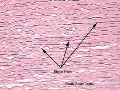
                 (B)yellow fibrous/ elastic tissue-thin and branched and made up of elastin protein

    FIBROUS TISSUE

    * these connective tissue contains fibroblast cells and animal's fibre
    *fibrous connective tissue classified into two groups-
    1. White fibrous connective tissue-it is made up of white fibre, e.g.- tendon.
    2. Yellow fibrous connective tissue- it is made up of yellow fibre. e.g.- ligament
    ADIPOSE TISSUE

    * it contains fat and fat forming cells.
    *it is further classified into two groups-
          White adipose tissue
          Brown adipose tissue.
    *in children, brown adipose tissue is present in excess amount , hence children less suffer from cold.
    *brown adipose tissue contain excess amount of mitochondria and very less amount of ATPs.
    *as age increases, brown adipose changes into white adipose tissue.
    *Blubber- it is thick pad of adipose tissue found in blue whale.
    *Hump-it is found in camel and ox, it is accumulation of adipose tissue.

    RETICULAR CONNECTIVE TISSUE

    *it is thin thread like structure of reticulin protein.
    *reticulin protein is generally store in lymphatic tissue. e.g. lymph, vermiform appendix, spleen.


    CONNECTIVE TISSUE

    *It helps in connection and binding of cells, tissue and different organ.
    *it helps in transportation of nutrients.
    *it gives mechanical support to body.
    *it also helps in transportation of hormones and enzymes.
    *it helps in hearing(malleus, incus, stapes)
    *it also helps in production of sound.
    *WHARTSON JELLY - an undifferentiated connective tissue, which is only found in umbilical cord of foetus.
    *MESENCHYME- undifferentiated connective tissue found everywhere except umbilical cord.
    *CLASSIFICATION OF CONNECTIVE TISSUE-


    Sunday, 21 February 2016

    STRATIFIED EPITHELIUM (COMPOUND EPITHELIUM)

    *cellls arranged in more than one layer.
    *basal layer of the tissue are either cuboidal or columnar , never squamous, so it is further classified into three types on the basis of cells present on its upper surface-

    1. Compound squamous epithelium/ pavement
    2. Compound columnar epithelium
    3. Compound squamous epithelium
    1. Compound squamous epithelium-this type of epithelium contains, squamous cells on free surface, it is further classified into two group on the basis if Keratin protein.

    #Compound keratinised squamous epithelium-it contains keratin protein, hence it is hard and dead. e.g.-epidermis of skin.

    #Compound non-keratinised squamous epithelium-it does not contain keratin protein, so it is relatively soft. it is present at all opening of the body which communicate with environment like mouth, anus ,nasal opening,ear opening, urinogenital opening.


    2. Compound cuboidal epithelium-this type of epithelium contains cuboidal cells on free surface.
    e.g.-testis, ovary

    3.Compound columnar epithelium-this type of epithelium contains columnar cells on its free surface.
    e.g.- epiglottis, gullet, larynx,pharynx,oesophagus(all parts are of food track).


    SPECIAL TYPE OF EPITHELIAL TISSUE

    A. Pseudo-stratified epithelium- cells arranged in single layer, but it apopears like arranged in more than one layer. e.g.-it is found near pharynx.

    B. Transitional epithelium-these epithelium do not contain basement membrane. in this type of epithelium cells are arranged in 4-5 layers, and cells of each layer have different function. e.g.- urethra, urinary bladder.

    Saturday, 20 February 2016

    SIMPLE EPITHELIAL TISSUE (EPITHELIUM)

    *first animal tissue found in coelentrata.
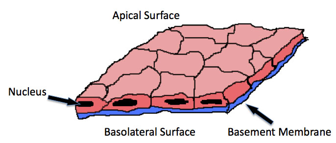
    *cells arranged in single or more than one layers, but the upper end of the layer is always free, means not covered.
    *presence of basement membrane, which is made up of gylcoprotein and polysaccharide.
    *epithelium divides throughout life.
    *cell of epithelium are avascular
    *Types of  epithelium on the basis of layer of the cell-

    1. Simple epithelium
    2. Compound/ Stratified epithelium
    1. Simple epithelium-cells are arranged in single layer. it is further classified into three different group-

    • Simple squamous epithelium/ Pavement
    • Simple cuboidal epithelium/ Germinal epithelium
    • Simple columnar epithelium
    Simple squamous epithelium- cells are arranged like tiles or scales of fish.
    -it contain very less quantity of matrix.
    -cells are smaller in size.
    -nucleus is at center of the cell.
    -distribution- (a) Pericardium (covering of heart)- parietal pericardium, visceral pericardium, pericardial fluid.
                          (b)Bowman's capsule-it is a part of nephron in kidney.
                          (c)Pleura of lungs- it is covering.

    Simple cuboidal epithelium- it contain very less quantity of matrix.
    -it is also known as germinal epithelium.
    -thyroid follicle of thyroid gland are made up of cuboidal epithelium.

    Simple columnar epithelium- cells are column like or row like.
    -nucleus present towards the base.
    -it contain less quantity of matrix.

    Modification of simple columnar epithelium
    (1) Ciliated epithelium- presence of small hairy structure on free surface of the cell, called cilia.
    -it performs specialised function.
    -it present in neck of nephron.
    -cilia of nephron helps in movement of filtrate from anterior end to posterior end.
    - it is also found in alveoli of lungs.
    -it also conduct egg in fallopian tube.

    (2)Sensory epithelium- cells of columnar epithelium modified to receive stimulus.
    -it is found in taste bud of tongue.
    -it is also found in organ of corti.
    -also present in rod and cones of retina of eye.
    -schnederian membrane of nasal tube.

    (3)Glandular epithelium- cells of columnar epithelium modified to form glands and gland specialised for secretion of substance.
                   Types of glands on the basis of number of cells present in gland.-
    #Unicellular glands- these gland contains only one cell. e.g.- goblet cell( found in intestine of human and secretes mucus), ebner's gland(present in tongue and secretes lipid digestive enzyme).

    #Multicellular glands-these gland contain more than one cells. e.g.- salivary gland, ovary, testis, liver, pancreas,thyroid, pituitary gland, lacrymal gland, mammary gland, sweat gland, parotid gland etc

                  Types of glands on the basis of transportation-
    #Exocrine gland-it has duct to transport their product. e.g.-salivary gland, lacrymal gland, mammary gland, liver etc.

    #Endocrine gland-these are duct less gland, they circulate their product through blood. e.g.- thyroid, pituitary,thymus etc.

    #Heterocrine gland-in this type of gland one part has duct and another is ductless. e.g.- pancreas.

                Types of glands on the basis of mode of secretion-
    #Holocrine gland-in this type of gland their total cytoplasm releases in the form of secretory material.e.g- sebaceous gland.

    #Apocrine gland-in this type of gland, small amount of cytoplasm releases in the form of secretory product. e.g.-mammary gland.

    #Merocrine gland-in this type of gland the secretory product releases through diffusion.e.g.- mammary gland.


    ANIMAL TISSUES, INTRODUCTION

    *group of cells having similar origin and function but their structure may or may not be similar , is commonly called TISSUE.
    *study of tissue is called Histology.
    *father and founder of Histology - Marcello Malpighi
    *Matrix- it is secretory product of cell in tissue. The quantity of matrix and chemical composition varies in different cell.
    *first of all tissue appeared in animals of phylum coelentrata.
    *on the basis of function tissues are classified into four types-
    1. Epithelial tissue- FUNCTION- absorption, secretion, protection etc.
    2. Connective tissue- FUNCTION- connection, binding, joining, attachment etc.
    3. Muscular tissue- FUNCTION- contraction and relaxation, movement and locomotion.
    4. Nervous tissue- FUNCTION- to receive stimulus and transport the transport the stimuli from one place to another.


    Wednesday, 17 February 2016

    MAMMALIA CLASS, VERTEBRATA.

    *these animals are warm blooded, hairy and have mammary or milk producing glands.
    *there are about 4.000 species of mammals found in world.
    *they are the only animals which nourish their young once with milk.
    *sebaceous glands(oil gland) and sudoriferous glands(sweat gland) are present in the skin.
    *teeth are heterodont and thecodont(embedded in the shocket of jaws). they develop twice in a life hence it is diphyodont
    *they possess a muscular diaphragm dividing trunk into thorax and abdomen.
    *mammals possess seven cervical vertebrae, except few.
    *the skull is dicondylic.
    *the heart is four chambered.
    *the coelom is divided into four cavities- a pericardial cavity, two pleural cavities and an abdominal cavity.
    *12 pair of cranial nerve.
    *each consists of three parts,external, middle and internal. pinna is a part of external ear
    *a copulatory organ is always present.
    *fertilization is internal.
    *except egg laying animal they are viviparous.
    *sub-class are Prototheria and Theria
    *e.g.-kangaroo, opossum, human etc
    OPOSSUM


    HUMAN


    KANGAROO


    Tuesday, 16 February 2016

    AVES-CLASS, VERTEBRATA

    *about 9000 species of aves are found
    *they are warm blooded animal.
    *they have bipeadal feather.
    *their fore-limbs are modified into wings.
    *the hind limbs are adapted for perching, walking or swimming.
    *only preen gland(oil gland) present on tail, and no other gland present on skin.
    *Ostrich and parrot lack oil glands
    *upper and lower jaws modified into beak,which lack teeth
    *legs bear horny epidermal skin.
    *respiration occurs in lungs.
    *syrinx produces voice in birds not larynx.
    *heart is four chambered, sinus venosus is absent.
    *renal portal system is very much reduced.
    *kidney is metanephric, which directly drain uric acid into cloca through ureter. urinary bladder is absent
    *Reha americana (South American Ostrich ) is the only bird which has urinary bladder.
    *cerebrum, cerebellum and optic lobes are developed organ of brain.
    *birds have 12 pair of cranial nerves.
    *bones are pneumatic,means they have air cavity to reduce weight , which helps in flying.
    *skull is monocondylic.
    *female has well developed single left ovary and oviduct.if right ovary and oviduct are present then they are non-functional.
    *sexual dimorphism is found in many birds.
    *fertilization is internal
    *birds are oviparous, means they lay eggs.
    *e.g.-Ostrich, Kiwi. Penguins, Parrot, Duck

    OSTRICH


    KIWI


    PENGUINS

    Friday, 12 February 2016

    REPTILIA CLASS, VERTEBRATA

    *they are about 6000 species
    *reptilia are creeping and burrowing cold blooded animals.
    *they bear scales
    *skin is dry, rough and without scales.
    *respiration is by lungs.
    *skull is monocondylic.
    *except in snake there are two pair of pentadactyl limbs, each with 5 digits bearing claws.
    *heart bear two auricles and partly divided ventricles
    *kidneys are metanephric
    *12 pairs of cranial nerves are present
    *ear consists of three parts- internal, middle anr wxternal.
    *snake do not possess ear.
    *all reptiles are carnivorous/insectivorous, only tortoise feed entirely on vegetation.
    *a typical cloaca is present.
    *fertilization is internal
    *e.g-Chelone(turtle), Testudo(tortoise), Chameleon(tree lizard),Naja(cobra),Bangarus(karait) etc

    TESTUDO


    CHELONE


    NAJA